
SPASTICITY
-
Spasticity also known as “Hypertonia” is a condition in which there is an abnormal increase in muscle tone and leads to muscle stiffness and rigidity. It makes mobility, movement and speech difficult.
Spasticity is a neurological disorder due to the damage in the nerve pathway (typically at the level of the brain or spinal cord). Spasticity is usually seen in individuals with spinal cord injury, multiple sclerosis, cerebral palsy, stroke, brain or head trauma, amyotrophic lateral sclerosis…
Among others, pharmacological treatments of spasticity include:
 Chemodenervation by intramuscular Botulinum toxin or its derivatives
Neurolysis with Phenol or ethanol injection
Conduction block with anesthetics or curare-like compounds
Chemodenervation by intramuscular Botulinum toxin or its derivatives
Neurolysis with Phenol or ethanol injection
Conduction block with anesthetics or curare-like compounds
-
Chemodenervation induced by intramuscular Botulinum toxin
Chemodenervation is performed when spasticity is severe, and the affected muscle is in specific locations. It consists of injecting Botulinum neurotoxins (Botox,® Myobloc,® Dysport,® and Xeomin®) into the spastic muscle to disrupt the contraction and allow muscle relaxation via inhibition of the exocytosis of synaptic vesicles containing acetylcholine. Among other uses, they are used for the treatment of children with cerebral palsy.
Neurofit has implemented an efficient and cost-effective model / assay to evaluate the anti-spastic potential of drug/compound treatments. The approach consists of measuring the depression of muscle electrical response (Compound Muscular Action Potential, CMAP) following the anti-spastic treatment. The CMAP measure is a clinically-relevant diagnostic tool for the evaluation of neuromuscular function.
Neurofit is able to characterize, evaluate and follow the impact of chemodenervation agents in rat or mice. The main and sensitive read-out is the measure of the amplitude of the compound muscular action potential (CMAP) in the injected muscle following the stimulation of the motor nerve. This electrical response is markedly reduced by intramuscular injection of Botulinum toxins and thus reflects the degree of muscle relaxation. The effect can be monitored up 120 days post-injection.
In addition to the measure of CMAP, there is a reduced weight bearing in the toxin-injected limb. Thus, the measure of weight bearing imbalance between the injected and the contralateral limb is another means to measure the impact of treatment on the muscle function.
-

Graphs and Data are from Toxicon X.2020 May23;7:100041. doi:10.1016/j. toxcx. 2020.100041 Cornet et al., Toxicon X. 2020 Sep; 7: 100041.
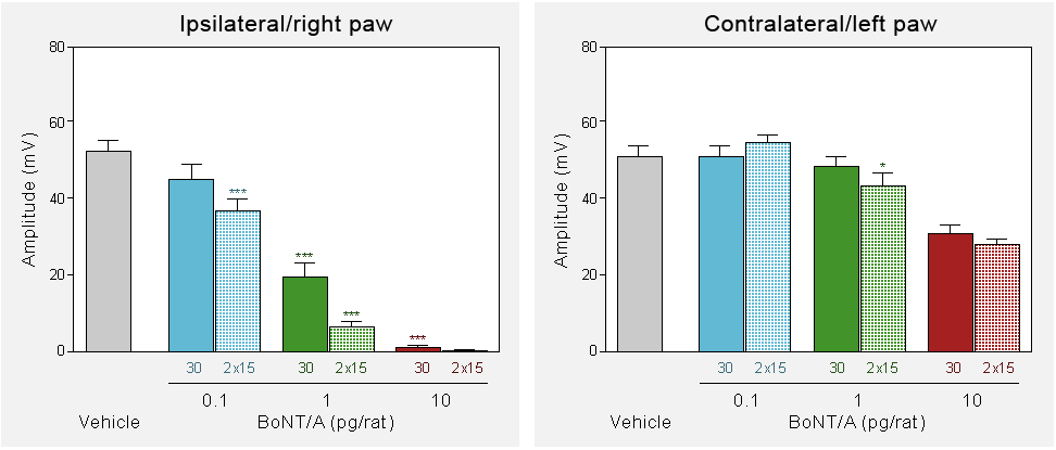
Intramuscular injection of Botulinum Toxin Type A (BoNT-A: 0.1, 1, or 10 pg/rat) into the right gastrocnemius lateralis of rats induces a dose-dependent depression of its amplitude of Compound Muscular Action potential (CMAP) as compared to the level recorded in the vehicle (Veh) injected rats. BoNT-A is administered into the head of the gastrocnemius lateralis muscle as a single bolus (30 μL), or into the heads of the gastrocnemius lateralis and gastrocnemius medialis as two 15 μL boluses. The effect at the contralateral side allows to assess the remote spread of BoNT-A.
*p < 0.05, ***p < 0.001 compared to the vehicle group. 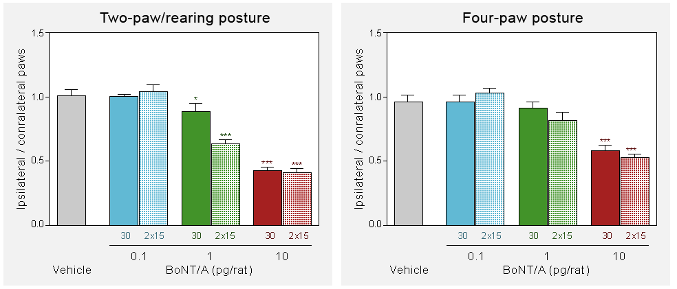
-
Intramuscular injection of Botulinum Toxin Type A (BoNT-A: 0.1, 1, or 10 pg/rat) into the right gastrocnemius lateralis of rats induces a dose-dependent decrease in the ratio of weight bearing between the ipsilateral and contralateral hindpaws as compared to the level recorded in the vehicle (Veh) injected rats. This weight bearing imbalance is observed in different posture of the rats. BoNT-A is administered into the head of the gastrocnemius lateralis muscle as a single bolus (30 μL), or into the heads of the gastrocnemius lateralis and gastrocnemius medialis as two 15 μL boluses. The effect at the contralateral side allows to assess the remote spread of BoNT-A.
*p < 0.05, ***p < 0.001 compared to the vehicle group.
-
Chemical Neurolysis
-
Neurolysis is the temporary denervation of a muscle or a group of muscles through perineural infiltration of chemicals causing non-selective denaturation of proteins of axons Neurolysis is most often performed with phenol and ethanol but hypertonic saline, glycerol, ammonium salt solutions, and chlorocresol have also been used in the past.
Neurofit is able to characterize, evaluate and follow the impact of neurolytic agents, such as Phenol, in the rat. The procedure involves a perineural injection of Phenol (or other neurolytic agents) onto the sciatic nerve and the evaluation of the amplitude of the compound muscular action potential (CMAP) in the gastrocnemius (target muscle) following the stimulation of the sciatic nerve. This electrical response is markedly reduced by the Phenol infiltration and thus reflects the degree of muscle relaxation. The effect of Phenol is very fast (within minutes) and can last for few months.
-
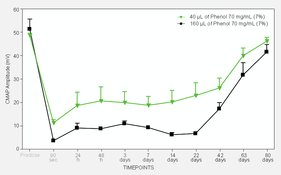
Perineural injection of 7% Phenol at the level of the sciatic nerve of rats induces a dose-dependent depression of the amplitude of Compound Muscular Action potential (CMAP) of the gastrocnemius. Hence, one single injection induces a very rapid (within 90 sec) marked depression of the muscle contraction and the effect lasts over several weeks before a clear sign of recovery is observed. A couple of months is needed for a full recovery. The treatment can be repeated to allow a continuous depression of the muscle contraction.
-
Non-destructive nerve block
-
The use of curare or curare-like agents as a non-destructive nerve block methods is common practice in anesthesiology to partially or completely block the motor nerve activity and consequently induces muscle relaxation. The first report to suggest the use of curare-like agents for the treatment of spasticity was published around 1945. However, the extreme toxicity of these agents has limited their use in the context of spasticity which requires a long-term follow up treatment.
-
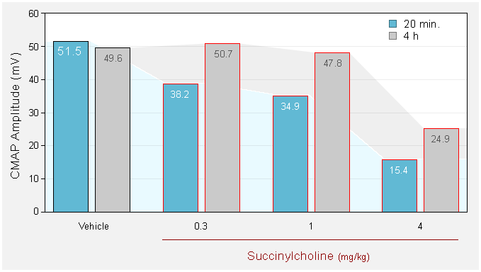
Intramuscular (gastrocnemius) injection of curare-like agent (succinylcholine) induces a dose-dependent depression of depression of the amplitude of Compound Muscular Action potential (CMAP) of the gastrocnemius. The depression of the muscle response is quick but the effect lasts for over few hours only.
You could also be interested in
-
SNCV
Sensory nerve conduction velocity is used in the diagnosis of nerve damage or nerve dysfunction.
CMAP
Compound muscle action potential consist in stimulating a nerve fiber and monitoring the response in the muscle.
-
Peripheral neuropathy
Result of damages of the peripheral nervous system. These troubles can be caused either by a trauma or diseases as for example diabetes.
STZ (peripheral neuropathy)
Diabetic neuropathy can be studied in rat following streptozotocin administration which induces a rapid and persistent hyperglycaemia.